2.1.7
Microscopes
Test your knowledge with free interactive questions on Seneca — used by over 10 million students.
Microscopes
It is important to understand the principles and limitations of optical microscopes, transmission electron microscopes and scanning electron microscopes.
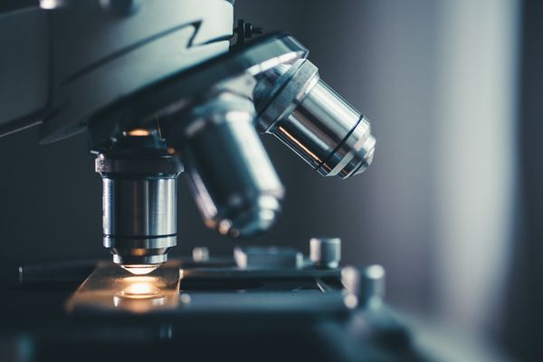
Optical (light) microscopes
- Visible light passes and is bent through the lens system to enable the user to see the specimen.
- The specimen can be alive.
- Individual cells are generally transparent and their components are not distinguishable unless they are coloured with special stains.
- Staining usually kills the cells.

Uses of light microscopes
- Most student microscopes are classified as light microscopes.
- Maximum resolution is 0.2 micrometres.
- The nucleus and mitochondria can be seen with a light microscope.
- The maximum magnification is around x1,500.

Electron microscopes
- In contrast to light microscopes, electron microscopes use a beam of electrons instead of a beam of light.
- This allows higher magnification and higher resolving power.
- This means that more detail can be seen.

Uses of electron microscopes
- Electron microscopes have a maximum resolution of 0.0002 micrometres.
- This is around 1000 times more than light microscopes.
- The maximum magnification is around x1,500,000.
Types of Electron Microscopes
There are two main types of electron microscopes: transmission (TEM) and scanning (SEM) electron microscopes.
TEM
- In a TEM, the electron beam penetrates the cell and provides details of a cell’s internal structures.
- TEMs use electromagnets to focus the electron beam.
- TEMs are high resolution microscopes.
- In thin specimens, you can see the internal structures of organelles such as chloroplasts.
SEM
- In a SEM, a beam of electrons moves back and forth across a cell’s surface, creating details of cell surface characteristics.
- SEMs knock electrons off the specimen and these electrons come together to form an image.
- SEM images can be three-dimensional.
- Specimens do NOT have to be thin like when using a TEM.
- Resolution is lower than that produced by a TEM.
1Biological Molecules
1.1Monomers & Polymers
1.2Carbohydrates
1.3Lipids
1.4Proteins
1.4.1The Peptide Chain1.4.2Investigating Proteins1.4.3Primary & Secondary Protein Structure1.4.4Tertiary & Quaternary Protein Structure1.4.5Enzymes1.4.6Factors Affecting Enzyme Activity1.4.7Enzyme-Controlled Reactions1.4.8End of Topic Test - Lipids & Proteins1.4.9A-A* (AO3/4) - Enzymes1.4.10A-A* (AO3/4) - Proteins1.4.11Diagnostic Misconceptions - Enzyme Inhibitors
1.5Nucleic Acids
1.6ATP
1.7Water
1.8Inorganic Ions
2Cells
2.1Cell Structure
2.2Mitosis & Cancer
2.3Transport Across Cell Membrane
2.4Cell Recognition & the Immune System
2.4.1Immune System2.4.2Phagocytosis2.4.3T Lymphocytes2.4.4B Lymphocytes2.4.5Antibodies2.4.6Primary & Secondary Response2.4.7Vaccines2.4.8HIV2.4.9Ethical Issues2.4.10End of Topic Test - Immune System2.4.11Exam-Style Question - Immune System2.4.12A-A* (AO3/4) - Immune System2.4.13Diagnostic Misconceptions - Humoral vs Cellular
3Substance Exchange
3.1Surface Area to Volume Ratio
3.2Gas Exchange
3.3Digestion & Absorption
3.4Mass Transport
3.4.1Haemoglobin3.4.2Oxygen Transport3.4.3The Circulatory System3.4.4The Heart3.4.5Blood Vessels3.4.6Cardiovascular Disease3.4.7Heart Dissection3.4.8Xylem3.4.9Phloem3.4.10Investigating Plant Transport3.4.11End of Topic Test - Mass Transport3.4.12A-A* (AO3/4) - Mass Transport3.4.13Diagnostic Misconceptions - Concentration Gradient3.4.14Diagnostic Misconceptions - Cardiac Cycle3.4.15Diagnostic Misconceptions - Carrying Capacity3.4.16Diagnostic Misconceptions - Translocation
4Genetic Information & Variation
4.1DNA, Genes & Chromosomes
4.2DNA & Protein Synthesis
4.3Mutations & Meiosis
4.4Genetic Diversity & Adaptation
4.5Species & Taxonomy
4.6Biodiversity Within a Community
4.7Investigating Diversity
5Energy Transfers (A2 only)
5.1Photosynthesis
5.1.1Overview of Photosynthesis5.1.2Photoionisation of Chlorophyll5.1.3Production of ATP & Reduced NADP5.1.4Cyclic Photophosphorylation5.1.5Light-Independent Reaction5.1.6A-A* (AO3/4) - Photosynthesis Reactions5.1.7Limiting Factors5.1.8Photosynthesis Experiments5.1.9End of Topic Test - Photosynthesis5.1.10A-A* (AO3/4) - Photosynthesis
5.2Respiration
5.2.1Overview of Respiration5.2.2Anaerobic Respiration5.2.3A-A* (AO3/4) - Anaerobic Respiration5.2.4The Link Reaction5.2.5The Krebs Cycle5.2.6Oxidative Phosphorylation5.2.7Respiration Experiments5.2.8End of Topic Test - Respiration5.2.9A-A* (AO3/4) - Respiration5.2.10Diagnostic Misconceptions - Aerobic vs Anaerobic
5.3Energy & Ecosystems
6Responding to Change (A2 only)
6.1Nervous Communication
6.2Nervous Coordination
6.3Muscle Contraction
6.4Homeostasis
6.4.1Overview of Homeostasis6.4.2Blood Glucose Concentration6.4.3Controlling Blood Glucose Concentration6.4.4End of Topic Test - Blood Glucose6.4.5Primary & Secondary Messengers6.4.6Diabetes Mellitus6.4.7Measuring Glucose Concentration6.4.8Osmoregulation6.4.9Controlling Blood Water Potential6.4.10ADH6.4.11End of Topic Test - Diabetes & Osmoregulation6.4.12A-A* (AO3/4) - Homeostasis6.4.13Diagnostic Misconceptions - Effect of ADH
7Genetics & Ecosystems (A2 only)
7.1Genetics
7.2Populations
7.3Evolution
7.3.1Variation7.3.2Natural Selection & Evolution7.3.3End of Topic Test - Populations & Evolution7.3.4Types of Selection7.3.5Types of Selection Summary7.3.6Overview of Speciation7.3.7Causes of Speciation7.3.8Diversity7.3.9End of Topic Test - Selection & Speciation7.3.10A-A* (AO3/4) - Populations & Evolution7.3.11Diagnostic Misconceptions - Types of Speciation
8The Control of Gene Expression (A2 only)
8.1Mutation
8.2Gene Expression
8.2.1Stem Cells8.2.2Stem Cells in Disease8.2.3End of Topic Test - Mutation & Gene Epression8.2.4A-A* (AO3/4) - Mutation & Stem Cells8.2.5Regulating Transcription8.2.6Epigenetics8.2.7Epigenetics & Disease8.2.8Regulating Translation8.2.9Experimental Data8.2.10End of Topic Test - Transcription & Translation8.2.11Tumours8.2.12Correlations & Causes8.2.13Prevention & Treatment8.2.14End of Topic Test - Cancer8.2.15A-A* (AO3/4) - Gene Expression & Cancer
8.3Genome Projects
Jump to other topics
1Biological Molecules
1.1Monomers & Polymers
1.2Carbohydrates
1.3Lipids
1.4Proteins
1.4.1The Peptide Chain1.4.2Investigating Proteins1.4.3Primary & Secondary Protein Structure1.4.4Tertiary & Quaternary Protein Structure1.4.5Enzymes1.4.6Factors Affecting Enzyme Activity1.4.7Enzyme-Controlled Reactions1.4.8End of Topic Test - Lipids & Proteins1.4.9A-A* (AO3/4) - Enzymes1.4.10A-A* (AO3/4) - Proteins1.4.11Diagnostic Misconceptions - Enzyme Inhibitors
1.5Nucleic Acids
1.6ATP
1.7Water
1.8Inorganic Ions
2Cells
2.1Cell Structure
2.2Mitosis & Cancer
2.3Transport Across Cell Membrane
2.4Cell Recognition & the Immune System
2.4.1Immune System2.4.2Phagocytosis2.4.3T Lymphocytes2.4.4B Lymphocytes2.4.5Antibodies2.4.6Primary & Secondary Response2.4.7Vaccines2.4.8HIV2.4.9Ethical Issues2.4.10End of Topic Test - Immune System2.4.11Exam-Style Question - Immune System2.4.12A-A* (AO3/4) - Immune System2.4.13Diagnostic Misconceptions - Humoral vs Cellular
3Substance Exchange
3.1Surface Area to Volume Ratio
3.2Gas Exchange
3.3Digestion & Absorption
3.4Mass Transport
3.4.1Haemoglobin3.4.2Oxygen Transport3.4.3The Circulatory System3.4.4The Heart3.4.5Blood Vessels3.4.6Cardiovascular Disease3.4.7Heart Dissection3.4.8Xylem3.4.9Phloem3.4.10Investigating Plant Transport3.4.11End of Topic Test - Mass Transport3.4.12A-A* (AO3/4) - Mass Transport3.4.13Diagnostic Misconceptions - Concentration Gradient3.4.14Diagnostic Misconceptions - Cardiac Cycle3.4.15Diagnostic Misconceptions - Carrying Capacity3.4.16Diagnostic Misconceptions - Translocation
4Genetic Information & Variation
4.1DNA, Genes & Chromosomes
4.2DNA & Protein Synthesis
4.3Mutations & Meiosis
4.4Genetic Diversity & Adaptation
4.5Species & Taxonomy
4.6Biodiversity Within a Community
4.7Investigating Diversity
5Energy Transfers (A2 only)
5.1Photosynthesis
5.1.1Overview of Photosynthesis5.1.2Photoionisation of Chlorophyll5.1.3Production of ATP & Reduced NADP5.1.4Cyclic Photophosphorylation5.1.5Light-Independent Reaction5.1.6A-A* (AO3/4) - Photosynthesis Reactions5.1.7Limiting Factors5.1.8Photosynthesis Experiments5.1.9End of Topic Test - Photosynthesis5.1.10A-A* (AO3/4) - Photosynthesis
5.2Respiration
5.2.1Overview of Respiration5.2.2Anaerobic Respiration5.2.3A-A* (AO3/4) - Anaerobic Respiration5.2.4The Link Reaction5.2.5The Krebs Cycle5.2.6Oxidative Phosphorylation5.2.7Respiration Experiments5.2.8End of Topic Test - Respiration5.2.9A-A* (AO3/4) - Respiration5.2.10Diagnostic Misconceptions - Aerobic vs Anaerobic
5.3Energy & Ecosystems
6Responding to Change (A2 only)
6.1Nervous Communication
6.2Nervous Coordination
6.3Muscle Contraction
6.4Homeostasis
6.4.1Overview of Homeostasis6.4.2Blood Glucose Concentration6.4.3Controlling Blood Glucose Concentration6.4.4End of Topic Test - Blood Glucose6.4.5Primary & Secondary Messengers6.4.6Diabetes Mellitus6.4.7Measuring Glucose Concentration6.4.8Osmoregulation6.4.9Controlling Blood Water Potential6.4.10ADH6.4.11End of Topic Test - Diabetes & Osmoregulation6.4.12A-A* (AO3/4) - Homeostasis6.4.13Diagnostic Misconceptions - Effect of ADH
7Genetics & Ecosystems (A2 only)
7.1Genetics
7.2Populations
7.3Evolution
7.3.1Variation7.3.2Natural Selection & Evolution7.3.3End of Topic Test - Populations & Evolution7.3.4Types of Selection7.3.5Types of Selection Summary7.3.6Overview of Speciation7.3.7Causes of Speciation7.3.8Diversity7.3.9End of Topic Test - Selection & Speciation7.3.10A-A* (AO3/4) - Populations & Evolution7.3.11Diagnostic Misconceptions - Types of Speciation
8The Control of Gene Expression (A2 only)
8.1Mutation
8.2Gene Expression
8.2.1Stem Cells8.2.2Stem Cells in Disease8.2.3End of Topic Test - Mutation & Gene Epression8.2.4A-A* (AO3/4) - Mutation & Stem Cells8.2.5Regulating Transcription8.2.6Epigenetics8.2.7Epigenetics & Disease8.2.8Regulating Translation8.2.9Experimental Data8.2.10End of Topic Test - Transcription & Translation8.2.11Tumours8.2.12Correlations & Causes8.2.13Prevention & Treatment8.2.14End of Topic Test - Cancer8.2.15A-A* (AO3/4) - Gene Expression & Cancer
8.3Genome Projects
Practice questions on Microscopes
Can you answer these? Test yourself with free interactive practice on Seneca — used by over 10 million students.
- 1
- 2What are the two main types of electron microscopes?Fill in the list
- 3
- 4Which of the following does NOT apply to SEM? Multiple choice
Unlock your full potential with Seneca Premium
Unlimited access to 10,000+ open-ended exam questions
Mini-mock exams based on your study history
Unlock 800+ premium courses & e-books